Techniques and equipment
In the Blanford lab
Quartz crystal microbalance
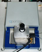 A quartz crystal microbalance has at its heart a resonant timing device like the one inside a quartz watch. On the surface of the crystal are two thin gold electrodes which drive the piezoelectric crystal at about 5 MHz. One of these faces acts as a sensor: the resonant frequency of the crystal drops proportionally to any increase in the mass of material adsorbed on the sensor surface. The precision of the frequency measurement is sufficiently high to measure mass changes in solution that correspond to less than 1% of a monolayer of a laccase-size (or BSA-size) protein. Our particular QCM, a q-sense E1, also has the ability to measure the energy dissipation (inversely proportional to the Q factor of the resonator) of the adsorbed layer. This additional information gives us a qualitative indication of the rigidity or stiffness of an adsorbed layer (partly related to how tightly it is attached) and allows us to model thickness and other layer properties when we move too far from the assumption built into the traditional Sauerbrey equation.
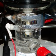
A third piece of information is obtained when the QCM cell is intergrated with protein film voltammetry measurements, allowing us to determine simultaneously the quantity of adsorbed protein, the rate at which it’s adsorbed, the quality of its mechanical coupling to the surface, the fraction of that protein that is oriented to allow rapid electron transfer between the electrode and protein.
Protein film voltammetry
 Protein Film Voltammetry (PFV), also sometimes known as Protein Film Electrochemistry (PFE), is a valuable technique for probing the reactions of redox-active proteins. The basic principle is the immobilisation of a (typically) sub-monolayer of protein molecules onto an electrode surface such that they retain their native activity. Electrode materials typically used are pyrolytic graphite, glassy carbon, and metals such as platinum and gold.
In a PFE experiment, electron flow, i.e., current, is recorded in response to an applied potential (driving force). The key advantage of PFE is the ability to precisely control the applied electrode potential, and thus intricate details in the behaviour of protein molecules can be revealed. In addition to the potential, the effect of changing experimental variables such as temperature, pH, and the composition and ionic strength of buffer solutions can be examined.
Redox catalysis (i.e., catalyic oxidation or reduction) by enzymes is especially suited to being probed using PFV. For example, O2 reduction to water requires four electrons and thus generates a negative catalytic current. By monitoring changes in the catalytic current as a function of the applied electrode potential, information about the catalytic behaviour of the enzyme can be deduced.
We use our two Ivium CompactStats for our routine DC electrochemical measurements and some of our impedance measurements.
Electrochemical impedance spectroscopy
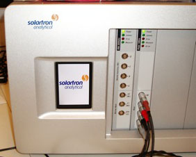 Impedance is the analogue to resistance when the current is changing with time, that is, it is the opposition to alternating current. As we scale up our electrode size, we will use the technique to measure ohmic resistance, activation polarisation resistance and transport properties. A related technique is Fourier transform voltammetry (FTV), which also allows us to explore rates of electron transfer and reaction kinetics. We use this technique to establish which modifications not only improve stability, but also speed up electron transfer to the enzyme and remove an electronic bottleneck. We use a Solartron ModuLab, shown at the right, for some of our EIS and FTV measurements.